
CPR Resource Center
The most comprehensive library of emergency training resources — including videos, articles, downloads, and more.
The most comprehensive library of emergency training resources — including videos, articles, downloads, and more.
An Electrocardiogram also known as an EKG or ECG is a medical test that measures the electrical activity of the heart. As the chambers of the heart receive these electrical impulses they contract and relax which produce specific waveforms that medical professionals can interpret. This data can ultimately tell medical professionals a few key things such as how fast, slow, or irregular a heart rhythm may be or if areas of the heart are too large, overworked and also provide early insight on if electrolyte values may be impaired.
An EKG can also analyze if there is a delayed or rapid conduction that takes place within the electrical pathways of the heart which produce different intervals such as the PR interval, the QRS interval, the QT interval, etc. While an EKG is a great tool to use for helping interpret the heart rhythm, it is subject to human error and there are several steps healthcare providers can take to ensure a proper EKG tracing is taken to include: good skin preparation and proper electrode placement.
Improper preparation could lead to an EKG that includes artifact. Artifact also known as artifactual signal is anything on an ECG that is not caused by the electrical currents generated by the heart which includes electrical interference, muscle tremors, patient movement, or loss of electrode contact. Most of these types of artifacts can be eliminated with proper skin prep and correct electrode placement.
Skin preparation prior to performing an EKG is extremely important. Did you know that an estimated 85-99% of cardiac monitoring or telemetry alarms are false? (Walsh-Irwin & Jurgens, 2015). Many times, EKGs or cardiac monitor readings pick up patient movement which could appear on screen to be life threatening arrythmias but the patient may have been moving around in bed, had a sudden cough, or slight enough moment to cause artifact. More times that not this is due to inadequate skin preparation.
This can lead to alarm fatigue, a false sense of security and uncertainty in cardiac monitoring which is a huge patient safety concern. Additionally, inadequate skin prep can result in increased healthcare costs. Depending on the brand, each electrode could cost upwards of 40 cents. That may not seem like a lot at first but those numbers can add up very quickly. Appropriate skin preparations need to be performed to ensure an accurate and reliable reading is present.
Appropriate skin prep includes shaving the hair at the electrode sides, cleaning each electrode site with soap and water or a non-alcohol-based wipe, drying the skin vigorously to improve blood flow, and using an abrasive prep pad or paper on the EKG electrode sides prior to placement.
Use of an abrasive prep pad or paper can reduce electrode impedance from 100,000 – 200,000 ohms to 1,000 – 5,000 ohms which drastically reduce the potential for motion “artifact” (Smith, 2016).
The next measure to improve EKGs is ensuring proper placement and positioning of the electrodes. All electrodes used when performing an EKG should be of the same brand. Additionally, you should avoid placing electrodes over bones or major muscles to eliminate any artifact. Try not to press directly in the center of the electrode as it can create air pockets from improper gel distribution that will also increase artifact. The most common type of EKG performed is a 3 lead ECG and a 12 lead EKG.
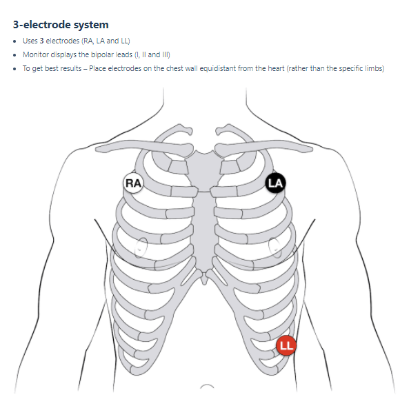
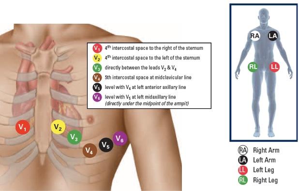
A 3 lead ECG is commonly used in Advanced Life Support settings. It is a quick and easy way to visualize a heart rhythm and provide care if indicated. For a 12 lead EKG, the process is a bit more extensive and electrode placement needs more thorough consideration. Although there are technically only 10 electrodes that are placed on the patient, there are 12 views of the heart that this test provides.
The correct placement for a 3 lead ECG monitoring and a 12 lead EKG are as followed:
EKGs are a great tool for trained healthcare professionals to use to identify abnormal heart rhythms and aid in diagnoses. EKGs should only be performed with a doctor’s order and by trained healthcare professionals. While most EKGs are performed in a routine outpatient setting, some may need to be performed rapidly as in situations where Advanced Life Support measures are being performed.
It is imperative to ensure proper skin preparation and electrode placement to allow healthcare professionals to provide reliable EKG tracings and rule out abnormalities or render treatment. So, if you are a healthcare professional trained in using EKG’s or cardiac monitoring, remember that these simple steps can improve the quality of your EKGs and lead to improved outcomes.
For more content from Code One Training Solutions follow us on Facebook, Instagram, LinkedIn or check out or Learning Center.
Electrocardiogram (ECG or EKG). (n.d.). Retrieved March 21, 2021, from https://www.heart.org/en/health-topics/heart-attack/diagnosing-a-heart-attack/electrocardiogram-ecg-or-ekg
Quick tip: How to take the perfect 12-lead ECG. (2015, July 05). Retrieved March 21, 2021, from https://www.emergencymedicinekenya.org/quick-tip-how-to-take-the-perfect-12-lead-ecg/
Phillips (n.d.). Improving ECG Quality. Retrieved March 21, 2021, from https://philipsproductcontent.blob.core.windows.net/assets/20170523/f2fc03ac224d4d5bb6aaa77c0151ac70.pdf
Smith, M. (2016, January 25). Rx for ECG Artifact. Retrieved March 21, 2021, from https://general-devices.com/rx-for-ecg-artifact/
Walsh-Irwin, Colleen & Jurgens, Corrine. (2015). Proper Skin Preparation and Electrode Placement Decreases Alarms on a Telemetry Unit. Dimensions of critical care nursing : DCCN. 34. 134-139. 10.1097/DCC.0000000000000108.
Help Me Find a Course